
Aug
14
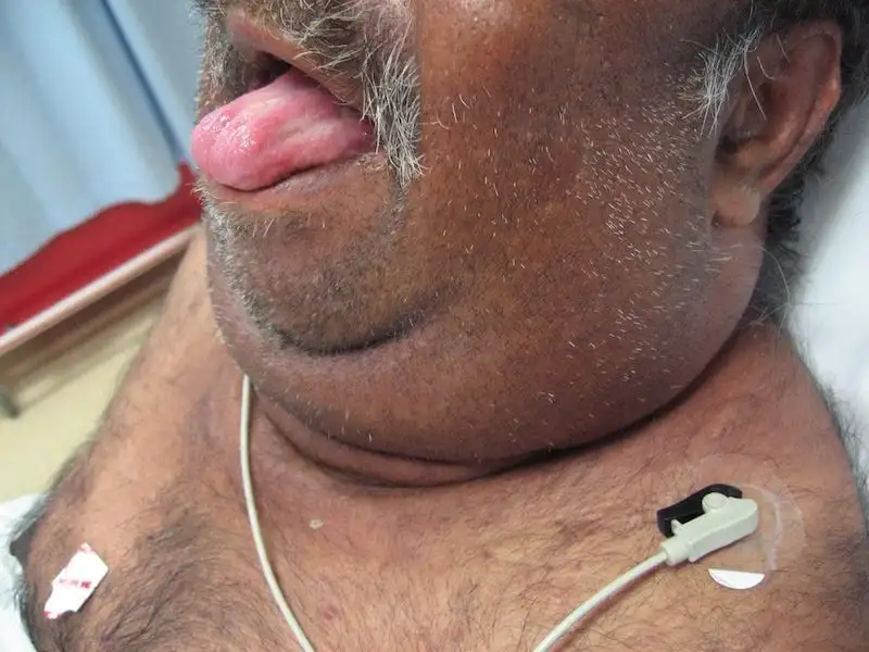
Category: Dental — Oral Surgery & Pathology | Medical Specialist: Oral and maxillofacial surgeon; oral medicine/oral pathology specialist; dentist | References: AAOMS; ADA; NIDCR Introduction: When a Tooth Infection Becomes an Emergency A toothache may seem like a problem that can wait until tomorrow. But when a dental infection begins spreading beneath the tongue and into the neck, the situation can change rapidly. Ludwig’s Angina is a rare but potentially life-threatening bacterial infection involving the floor of the mouth and the spaces beneath the jaw. Unlike a typical dental abscess, it can spread through interconnected tissue spaces and push the tongue upward, threatening the airway. That is what makes Ludwig’s Angina particularly important for both patients and dental professionals: the biggest danger isn’t simply the infection—it is the swelling that can interfere with breathing. Modern antibiotics, advanced imaging and better airway management have dramatically improved outcomes. Yet the condition still requires urgent medical attention because deterioration can occur quickly. Emergency warning: Difficulty breathing or swallowing, drooling, rapidly increasing swelling under the jaw or tongue, or a muffled/altered voice should be treated as an emergency. Do not wait for a routine dental appointment. What Is Ludwig’s Angina? Ludwig’s Angina is a rapidly spreading cellulitis, meaning inflammation and infection of soft tissues rather than a simple, well-contained abscess. It classically involves three connected anatomical spaces: The infection can cause firm swelling of these tissues and elevate the tongue. Because the tissues around the airway have limited room to expand, swelling can eventually make breathing difficult. The condition was first described in 1836 by German physician Wilhelm Friedrich von Ludwig. Despite its name, Ludwig’s Angina has nothing to do with the heart condition commonly called angina. What Causes Ludwig’s Angina? Dental infections are the leading cause One of the most important insights about Ludwig’s Angina is that a serious neck infection can begin with something as ordinary as an untreated dental infection. In particular, infections involving the lower second and third molars are strongly associated with Ludwig’s Angina. Current clinical references report that odontogenic infections account for the large majority of cases. Why do these teeth matter? The roots of lower molars can lie close to, or below, the attachment of the mylohyoid muscle. This anatomical relationship provides a pathway through which infection can spread into the submandibular tissues. Common dental sources include: Other possible causes Although dental infections dominate, Ludwig’s Angina can occasionally develop after: The infection is usually polymicrobial, meaning several types of bacteria may be involved. Aerobic and anaerobic organisms—including Streptococcus, Staphylococcus and anaerobic bacteria such as Bacteroides—can contribute. Who Is at Higher Risk? Ludwig’s Angina can occur in otherwise healthy people, but certain factors can increase susceptibility or make infection more difficult to control. Risk factors include: This is one reason preventive dental care matters beyond protecting teeth. Treating an infected tooth early can sometimes prevent a localized dental problem from becoming a deep neck infection. Ludwig’s Angina Symptoms: What Should You Watch For? Early symptoms can resemble an ordinary dental infection, which makes recognition especially important. Common symptoms Patients may experience: A characteristic finding is firm, bilateral swelling around the submandibular and submental areas, sometimes described as a “bull neck” appearance. The red flags matter most Not every symptom carries the same level of urgency. Symptom/sign What it may indicate Urgency Toothache with localized swelling Dental infection Prompt dental assessment Fever + jaw/neck swelling Spreading infection Urgent medical assessment Difficulty swallowing Increasing floor-of-mouth swelling Emergency Drooling Difficulty managing secretions Emergency Tongue elevation/swelling Airway displacement Emergency Muffled or altered voice Possible upper-airway involvement Emergency Difficulty breathing or stridor Threatened airway Immediate emergency care The evidence-based approach is straightforward: airway concerns take priority over diagnostic imaging or routine dental treatment. Why Ludwig’s Angina Can Become Dangerous So Quickly The anatomy of the floor of the mouth explains much of the danger. Think of the neck as a series of connected compartments. An infection does not necessarily remain where it started. It can travel along tissue planes, causing inflammation and swelling in neighboring spaces. As swelling increases, the tongue may be pushed upward and backward. The surrounding tissues can also become increasingly stiff and swollen. This creates a dangerous sequence: Dental infection → tissue-space spread → floor-of-mouth swelling → tongue elevation → airway narrowing → breathing difficulty That progression is why Ludwig’s Angina is considered a medical emergency rather than simply a severe dental infection. Ludwig’s Angina vs. a Typical Dental Abscess These conditions can start from the same dental problem, but their clinical behavior is different. Feature Typical Dental Abscess Ludwig’s Angina Main location Tooth/gum or localized tissue Floor of mouth and submandibular spaces Swelling Usually localized Often firm and more diffuse Airway risk Usually low Potentially life-threatening Tongue elevation Uncommon Characteristic concern Difficulty swallowing May occur Common Breathing difficulty Uncommon Major warning sign Treatment Dental source control ± antibiotics Airway management + IV antibiotics ± surgery The key lesson is that a dental infection should never be judged only by how painful the tooth feels. The location and direction of swelling can be much more important. How Is Ludwig’s Angina Diagnosed? Diagnosis is primarily clinical. A healthcare professional examines the mouth, tongue, jaw and neck while assessing the patient’s ability to breathe and manage secretions. Imaging A contrast-enhanced CT scan of the neck can help determine how far the infection has spread and whether an abscess has developed. However, imaging should not delay airway management in someone whose breathing is threatened. A patient who cannot safely lie flat or manage secretions may need immediate airway intervention rather than being sent for a scan first. Ultrasound may also be useful in selected situations, particularly when clinicians need additional information without immediately sending an unstable patient for CT. Ludwig’s Angina Treatment Treatment is typically carried out in a hospital and involves several teams, often including emergency medicine, anesthesiology, oral and maxillofacial surgery, ENT and critical care. 1. Protect the airway first This is the most important principle. If swelling threatens