Category: Dental — Oral Medicine & Mucosal Conditions | Medical Specialist: Dentist; oral medicine specialist; dermatologist or physician when indicated | References: AAOM; ADA; NIDCR
Introduction: When a “White Patch” Is More Than a White Patch

A white, lace-like pattern inside the cheek can look harmless. Sometimes it is. But when those white lines persist, become sore, or appear alongside red, raw areas, they may point to a condition that deserves professional attention.
Oral Lichen Planus (OLP) is a chronic inflammatory condition affecting the tissues inside the mouth. It can be painless and easy to overlook—or painful enough to make eating, brushing, and even speaking uncomfortable. The challenge is that it can resemble several other oral conditions, which is why recognizing the pattern is only the first step.
The good news? Oral lichen planus can usually be managed. The goal is not simply to make a lesion “disappear,” but to control inflammation, relieve symptoms, identify possible triggers, protect oral health, and monitor persistent or changing areas over time.
Important: Oral lichen planus should be diagnosed by a qualified dentist, oral medicine specialist, dermatologist, or physician. A persistent mouth lesion should not be self-diagnosed.
What Is Oral Lichen Planus?
Oral lichen planus is a chronic inflammatory disorder involving the oral mucosa. It is generally considered an immune-mediated condition in which immune cells, particularly cytotoxic T cells, attack cells within the oral epithelium. However, the exact trigger behind this immune response remains uncertain.
Unlike a contagious infection, oral lichen planus cannot be passed from one person to another. It may occur on its own or alongside lichen planus affecting the skin or other mucosal surfaces. Interestingly, the mouth can sometimes be the only affected area.
Common Areas Affected
OLP may involve:
- Inner cheeks
- Tongue
- Gums
- Lips
- Roof or floor of the mouth
The inside of the cheeks is particularly characteristic, especially when lesions appear symmetrically.
What Causes Oral Lichen Planus?
This is one of the frustrating aspects of OLP: there is usually no single identifiable cause.
Researchers believe that an abnormal immune response plays the central role. Instead of protecting oral tissues, immune cells target epithelial cells, producing chronic inflammation.
Several factors may contribute to or resemble OLP:
1. Immune-System Activity
OLP is strongly associated with a T-cell-mediated inflammatory response. In many patients, however, the reason this response begins remains unknown.
2. Medications
Some medicines can produce oral lichenoid reactions, which can look remarkably similar to oral lichen planus.
Potentially associated medications include certain:
- Anti-inflammatory drugs
- Blood-pressure and cardiovascular medicines
- Antimalarial drugs
- Antibiotics
- Antiretroviral medicines
This distinction matters because a medication-related lichenoid reaction may improve after the responsible drug is identified and appropriately changed under medical supervision.
3. Dental Materials and Contact Reactions
In some cases, oral lichenoid lesions develop near dental restorations or in response to contact allergens. Materials such as metals, resins, or certain ingredients in oral-care products can occasionally be involved.
That does not mean every person with OLP needs dental fillings removed. Removing restorations without evidence of a relevant contact reaction is unlikely to cure OLP.
4. Stress and Local Trauma
Psychological stress and local mechanical irritation may aggravate existing lesions in some individuals. This is better understood as a potential trigger or exacerbating factor rather than a proven underlying cause.
5. Other Associations
Hepatitis C has been associated with lichen planus in some populations, although the strength of this relationship varies geographically. Your clinician can decide whether additional medical evaluation is appropriate based on your individual history.
Oral Lichen Planus Symptoms: What Does It Look and Feel Like?
OLP does not look the same in everyone. Some people have no discomfort at all, while others experience significant burning and soreness.
Common Signs and Symptoms
Look out for:
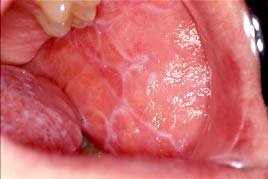
- White, web-like or lace-like lines
- White patches or plaques
- Red, inflamed areas
- Raw or eroded tissue
- Mouth ulcers
- Burning or stinging
- Gum redness and peeling
- Sensitivity to spicy, acidic, or salty foods
- Pain while eating or brushing
The classic white, branching lines are called Wickham striae. They are one of the visual clues clinicians may recognize during an oral examination.
The Erosive Form Can Be More Painful
Erosive oral lichen planus can produce red areas, superficial erosions, and ulceration. The gums may become particularly uncomfortable, sometimes producing a pattern known as desquamative gingivitis, where the surface tissue becomes red and peels away.
This is where OLP can stop being a cosmetic concern and become a quality-of-life issue.
Imagine trying to eat citrus fruit when the lining of your mouth is already inflamed. For someone with erosive OLP, that everyday experience can be surprisingly painful.
Oral Lichen Planus vs Other Common Mouth Lesions
Not every white or red oral lesion is OLP. Several conditions can look similar.
| Condition | Typical appearance | Pain | Key consideration |
|---|---|---|---|
| Oral Lichen Planus | Lacy white lines, plaques, red/erosive areas | Variable | Often chronic and may be symmetrical |
| Oral Thrush | Creamy white patches | Often burning | Usually associated with Candida infection |
| Leukoplakia | Persistent white patch | Often painless | Requires professional evaluation |
| Canker sore | Localized ulcer | Often painful | Usually heals within a limited period |
| Oral lichenoid reaction | Similar to OLP | Variable | May relate to medication or contact exposure |
| Oral cancer | Persistent ulcer, lump, red/white lesion or other change | May be painless | Requires prompt assessment |
The important takeaway is simple: appearance alone cannot always establish the diagnosis.
How Is Oral Lichen Planus Diagnosed?
Diagnosis usually begins with a detailed oral examination and medical history.
Your clinician may ask about:
- How long the lesion has been present
- Pain or burning
- Medication history
- Smoking and alcohol use
- Dental restorations
- Other skin or mucosal symptoms
- Previous episodes of similar lesions
Why Is a Biopsy Sometimes Necessary?
A biopsy may be recommended when the appearance is unusual, the diagnosis is uncertain, or an area needs to be evaluated for another condition.
Microscopic examination can support the diagnosis and help distinguish OLP from other lichenoid conditions. Biopsy can also be important when clinicians need to rule out dysplasia or oral cancer.
This is an important distinction: a biopsy does not mean your dentist thinks you have cancer. It is often a diagnostic safety step.
Oral Lichen Planus Treatment: What Actually Helps?
There is currently no guaranteed permanent cure for every case of OLP. Treatment is generally focused on controlling inflammation and symptoms while monitoring the condition.
1. Topical Corticosteroids
For symptomatic oral disease, topical corticosteroids are commonly used and are considered a mainstay of treatment.
They may be delivered as:
- Oral gels or pastes
- Rinses
- Sprays
- Other clinician-directed topical preparations
More severe disease may require additional therapies.
Because prolonged steroid use inside the mouth can increase the risk of oral candidiasis and other complications, treatment should be supervised by a healthcare professional.
2. Symptom Relief
Depending on the situation, clinicians may recommend treatments to reduce:
- Burning
- Pain
- Inflammation
- Oral sensitivity
Topical anesthetic preparations may sometimes provide temporary relief.
3. Treating Possible Triggers
If a medication or contact allergen appears responsible for a lichenoid reaction, the clinician may investigate whether the suspected trigger can be removed or replaced.
Never stop a prescription medication on your own. Any medication change should be made with the prescribing healthcare professional.
4. Systemic Treatment for Severe Disease
Severe or treatment-resistant cases may occasionally require systemic corticosteroids or other immunomodulatory medications.
Options reported in specialist management include agents such as methotrexate, mycophenolate, azathioprine, hydroxychloroquine, and others. These are not routine treatments for everyone with OLP and require specialist assessment because their risks and monitoring requirements vary.
Daily Oral Care: Small Habits That Make a Difference
Managing OLP isn’t only about medication.
Gentle, consistent oral care can make daily life considerably easier.
Practical Tips
- Brush twice daily with a soft toothbrush.
- Avoid aggressively brushing irritated tissues.
- Use an alcohol-free mouthwash if conventional mouthwash causes burning.
- Avoid foods that clearly trigger pain, particularly spicy, acidic, or salty foods.
- Consider a less-irritating toothpaste if conventional toothpaste causes discomfort.
- Maintain regular dental examinations.
- Avoid tobacco.
- Limit excessive alcohol consumption.
The NHS similarly recommends gentle oral hygiene and avoiding foods or alcohol that worsen mouth soreness.
One useful mindset is to think of your mouth as an environment that needs less irritation, not more intervention. When the tissue barrier is already inflamed, aggressive brushing, harsh mouthwashes, and unnecessary dental procedures can make symptoms worse.
Can Oral Lichen Planus Become Oral Cancer?
This is probably the question patients worry about most—and it deserves a balanced answer.
Research suggests that a small proportion of people diagnosed with OLP may develop oral squamous cell carcinoma. A 2024 systematic review and meta-analysis involving more than 38,000 patients estimated a pooled malignant transformation ratio of approximately 1.43%. Higher risk was associated with factors including epithelial dysplasia, tobacco and alcohol exposure, tongue involvement, and atrophic or erosive lesions.
That number should not be interpreted as “OLP causes cancer.” Most people with OLP do not develop oral cancer.
Instead, it explains why long-term professional monitoring matters.
Red Flags That Need Prompt Evaluation
Contact your dentist or oral medicine specialist if you notice:
- A persistent ulcer
- A lump or thickened area
- Bleeding without an obvious cause
- A lesion that is rapidly changing
- Increasing pain in one particular area
- A red or mixed red-and-white patch that persists
- Difficulty swallowing or persistent difficulty moving the tongue
Persistent or changing lesions may require examination and sometimes biopsy.
Oral Lichen Planus: Key Insights at a Glance
| Key insight | Why it matters |
| OLP is inflammatory and immune-mediated | It is not simply an infection |
| It can be painless | Absence of pain does not eliminate the need for evaluation |
| Erosive OLP can be painful | Treatment may focus heavily on symptom control |
| Similar-looking conditions exist | Diagnosis should not rely on photographs alone |
| Biopsy may be recommended | It can clarify diagnosis and exclude dysplasia/cancer |
| Long-term monitoring matters | A small malignant transformation risk has been reported |
| Oral hygiene remains important | Healthy gums reduce additional sources of inflammation |
| Tobacco avoidance is important | Tobacco is also associated with oral cancer risk |
Living With Oral Lichen Planus: A Long-Term Perspective
One of the biggest misconceptions about chronic oral conditions is that successful treatment always means making every visible mark disappear.
With OLP, success may look different.
It may mean eating comfortably again. It may mean fewer flare-ups, healthier gums, less burning, or simply knowing that a persistent lesion is being properly monitored.
Because oral lichen planus can last for years, management is often a long-term partnership between the patient and dental or medical team, rather than a one-time treatment.
And that is perhaps the most useful perspective: don’t panic when you hear the word “chronic,” but don’t ignore it either.
Frequently Asked Questions
Is oral lichen planus contagious?
No. Oral lichen planus is not considered a contagious infection and cannot ordinarily be spread through kissing, sharing utensils, or normal contact.
Can oral lichen planus go away?
Some symptoms may improve, but oral disease can persist for years and may fluctuate between periods of improvement and flare-ups.
Can stress cause oral lichen planus?
Stress may aggravate symptoms or contribute to flare-ups in some people, but it is not considered the sole underlying cause.
Should I have my dental fillings removed?
Not automatically. Dental materials can occasionally be associated with lichenoid reactions, but indiscriminate removal of restorations is not recommended. A clinician should determine whether there is evidence of a relevant contact reaction.
Is oral lichen planus cancer?
No. OLP is an inflammatory condition, not cancer. However, research indicates a small risk of malignant transformation, which is why persistent or changing lesions require appropriate follow-up.
Which specialist treats oral lichen planus?
Depending on your healthcare system, diagnosis and management may involve an oral medicine specialist, dentist, dermatologist, oral and maxillofacial specialist, or physician.
Final Thoughts
Oral Lichen Planus can be easy to overlook but shouldn’t be ignored. Its characteristic white patterns, red erosions, burning, and gum changes can affect everything from eating to everyday oral hygiene.
The smartest approach is neither alarm nor neglect. Get persistent lesions evaluated, understand what may trigger your symptoms, maintain gentle oral hygiene, follow your treatment plan, and attend recommended follow-up appointments.
Most importantly, don’t attempt to diagnose every white patch in your mouth from an online photograph. A professional examination—and when appropriate, a biopsy—provides far more reliable information.
Take the Next Step
If you have persistent white, red, sore, or ulcerated areas in your mouth, schedule an examination with your dentist or an oral medicine specialist. Early assessment can provide reassurance, appropriate treatment, and a clear monitoring plan.
For more educational content, explore related articles on gingivitis, periodontitis, oral ulcers, dental care, and oral cancer warning signs on your website.
Have you experienced recurring mouth soreness, burning, or unexplained white patches? Share your experience or questions in the comments—your story may help someone else recognize the importance of getting persistent oral changes checked.
Medical Disclaimer
This article is intended for general educational purposes only and does not replace professional medical or dental advice, diagnosis, or treatment. Medication names and treatment approaches should be discussed with a qualified healthcare professional. Any persistent, changing, bleeding, ulcerated, or suspicious oral lesion should be professionally evaluated.
Suggested Authoritative Resources
- DermNet: Oral Lichen Planus — clinical features, diagnosis, complications, and treatment.
- NHS: Lichen Planus — patient-friendly information on symptoms and oral self-care.
- PubMed: 2024 Systematic Review and Meta-analysis — evidence concerning malignant transformation risk.