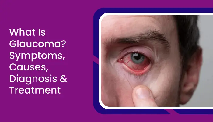
Category: Ophthalmology | Medical Specialist: Ophthalmologist; optometrist for appropriate conditions | References: American Academy of Ophthalmology (AAO); WHO; CDC Introduction: The Eye Disease You May Not Notice Imagine gradually losing part of your vision without realizing it. You can still read, recognize faces, and use your phone—yet your peripheral vision is quietly narrowing. That is one of the most troubling things about Glaucoma: in its most common forms, it can progress for years with few or no noticeable symptoms. By the time vision changes become obvious, permanent optic nerve damage may already have occurred. The National Eye Institute notes that many people with glaucoma do not know they have it because early disease often causes no symptoms. The good news is equally important: early detection and appropriate treatment can slow or stop further vision loss. This guide explains what glaucoma is, why it develops, which symptoms deserve attention, how doctors diagnose it, and how modern treatment can help preserve sight. What Is Glaucoma? Glaucoma is not a single disease but a group of eye conditions that damage the optic nerve, the structure responsible for carrying visual information from the eye to the brain. If the damage progresses, it can lead to irreversible vision loss or blindness. A useful way to understand the optic nerve is to think of it as a communication cable. The retina collects visual information, while the optic nerve transmits that information to the brain. Glaucoma gradually damages the nerve fibers within this pathway. Is glaucoma the same as high eye pressure? No. High intraocular pressure (IOP) is an important risk factor, but it does not automatically mean someone has glaucoma. Some people have elevated eye pressure without optic nerve damage, while others develop glaucoma despite having pressure within the range traditionally considered normal. This is why a pressure reading alone cannot diagnose glaucoma. Types of Glaucoma Different forms of glaucoma develop in different ways. Type What happens Typical pattern Primary open-angle glaucoma Fluid drains too slowly through the eye’s drainage system Usually gradual and painless Angle-closure glaucoma The drainage angle becomes blocked May develop suddenly and requires urgent treatment Normal-tension glaucoma Optic nerve damage occurs despite apparently normal eye pressure Often gradual Congenital glaucoma Abnormal development of the eye’s drainage system Usually appears during infancy or childhood Secondary glaucoma Develops because of another condition, medication, injury, or eye problem Depends on the underlying cause Primary open-angle glaucoma is the most common form in the United States, while angle-closure glaucoma is less common but can become an emergency. What Causes Glaucoma? There isn’t one universal cause. In many cases, the problem involves the way fluid called aqueous humor circulates and drains through the eye. When drainage is impaired, pressure may increase and contribute to optic nerve damage. However, the relationship between pressure and nerve damage is more complicated than simply saying “high pressure causes glaucoma.” Researchers continue to investigate why some optic nerves are more vulnerable than others. Important glaucoma risk factors Your risk may be higher if you have: Having a risk factor does not mean you will definitely develop glaucoma. It simply makes regular eye examinations more important. Glaucoma Symptoms: Why Early Detection Matters The most difficult thing about glaucoma is that the early stages can be almost invisible. Early symptoms For many people with open-angle glaucoma, there are no noticeable early symptoms. Vision can appear completely normal while optic nerve damage is occurring. As the condition progresses, people may develop: Peripheral vision may disappear gradually, which means the brain can adapt to changes without the person immediately recognizing what is happening. Acute angle-closure glaucoma: know the warning signs Angle-closure glaucoma can sometimes develop suddenly. Symptoms can include: These symptoms require urgent medical attention because acute angle closure can threaten vision quickly. How Is Glaucoma Diagnosed? A common misconception is that a quick “eye pressure test” is enough. It isn’t. A comprehensive eye examination may include several assessments that work together to determine whether the optic nerve is healthy. 1. Tonometry This measures intraocular pressure. Although useful, pressure is only one piece of the puzzle. A single normal pressure measurement does not rule out glaucoma. 2. Dilated eye examination The eye specialist examines the retina and optic nerve through a dilated pupil to look for structural signs of damage. 3. Visual field testing This checks how well you can see objects in different parts of your visual field. It can identify peripheral vision loss that you may not have noticed in everyday life. 4. Optic nerve imaging Modern eye clinics may use technologies such as optical coherence tomography (OCT) to measure and monitor the retinal nerve fiber layer and other structures. 5. Additional testing Depending on the situation, your ophthalmologist may evaluate the drainage angle, corneal thickness, optic nerve appearance, and changes over time. The key insight is simple: glaucoma diagnosis is based on a combination of structural and functional information, not one number. Glaucoma Treatment: Protecting the Vision You Still Have There is currently no cure that reverses established glaucomatous optic nerve damage. However, treatment can often slow or prevent additional damage. The central treatment goal is generally to reduce the risk of further optic nerve damage, commonly by lowering eye pressure. 1. Prescription eye drops Eye drops are among the most common treatments. Different medications work in different ways. Some increase fluid drainage, while others reduce the amount of fluid produced inside the eye. Prostaglandin analogues, beta blockers, alpha agonists, carbonic anhydrase inhibitors, and other medication classes may be used depending on the patient’s needs. One practical challenge is adherence. Glaucoma drops may not make you feel better because glaucoma often causes no symptoms. That can make it tempting to skip doses. But the purpose of treatment is not necessarily to improve today’s vision—it is to protect tomorrow’s vision. 2. Laser treatment Laser procedures can improve fluid drainage and lower eye pressure in appropriate patients. Depending on the type of glaucoma, procedures may include laser trabeculoplasty or other laser techniques. Laser treatment

Month: August 2026
Category: Orthopedics & Musculoskeletal | Medical Specialist: Orthopedic surgeon; rheumatologist; physiatrist; physiotherapist depending on condition | References: American Academy of Orthopaedic Surgeons (AAOS); American College of Rheumatology (ACR); WHO Introduction: When Everyday Movement Starts to Hurt A staircase you once climbed without thinking can suddenly feel like a challenge. Getting out of a chair may take an extra moment. Even opening a jar or taking a morning walk can become uncomfortable. These seemingly small changes can be signs of Osteoarthritis, the most common form of arthritis. Osteoarthritis is often described as a condition caused by “wear and tear.” But that description is misleading. The disease is more complex: it involves changes to cartilage, bone, the joint lining, ligaments, tendons, and surrounding muscles. And it is remarkably common. The World Health Organization estimates that about 528 million people worldwide were living with Osteoarthritis in 2019. The knee is the most frequently affected joint, followed by the hip and hand. The encouraging part? Osteoarthritis does not automatically mean giving up an active life. With the right combination of exercise, weight management, pain control, rehabilitation, and—when necessary—surgery, many people can maintain meaningful mobility and independence. What Is Osteoarthritis? Osteoarthritis is a long-term joint condition in which the tissues forming and supporting a joint gradually change. Healthy cartilage provides a smooth, resilient surface that helps bones move against one another. With Osteoarthritis, cartilage can deteriorate, while the underlying bone and other joint tissues may also undergo structural changes. Bone spurs, known as osteophytes, can develop around the joint. Commonly affected areas include: Importantly, Osteoarthritis is not simply an inevitable part of getting older. Age increases risk, but younger adults can also develop it, particularly after joint injuries, repetitive stress, or because of inherited joint abnormalities. Osteoarthritis vs Other Types of Arthritis Not all arthritis behaves the same way. Understanding the difference can help explain why treatment varies from person to person. Feature Osteoarthritis Rheumatoid Arthritis Main mechanism Structural changes and breakdown of joint tissues Autoimmune inflammation Typical pattern Often affects knees, hips, hands and spine Often affects multiple joints symmetrically Morning stiffness Usually brief, often under 30 minutes Frequently longer-lasting Common symptoms Pain with activity, stiffness, reduced movement Pain, swelling, stiffness and systemic symptoms Treatment focus Exercise, rehabilitation, weight management, symptom control Disease-modifying medicines and inflammation control This distinction matters because persistent joint pain should not automatically be labelled as Osteoarthritis. Conditions such as rheumatoid arthritis, gout, infection, or injuries can produce similar symptoms and may require very different treatment. What Causes Osteoarthritis? There is rarely one single cause. Instead, Osteoarthritis develops through an interaction between factors affecting the joint itself and factors affecting the body as a whole. 1. Age The risk increases with age because joints undergo biological and structural changes over time. However, ageing alone does not guarantee Osteoarthritis. 2. Previous Joint Injury A fracture, ligament injury, meniscus injury, or significant trauma can increase the likelihood of developing Osteoarthritis in that joint later. This is one reason that an apparently “old” injury can remain relevant years after recovery. 3. Excess Body Weight Weight can increase mechanical stress on weight-bearing joints such as the knees and hips. But the relationship is more complicated than simple pressure: metabolic and inflammatory processes associated with excess body weight may also contribute. 4. Repetitive Joint Stress Certain occupations and sporting activities can expose particular joints to repetitive loads. The risk depends on the joint, intensity, duration, biomechanics, and individual susceptibility. 5. Genetics Some people inherit characteristics that make their joints more vulnerable to Osteoarthritis. 6. Abnormal Joint Structure A joint that develops differently from normal may experience unusual mechanical forces, increasing the risk of later damage. Symptoms of Osteoarthritis Osteoarthritis often develops gradually, so the earliest warning signs can be easy to dismiss. Common symptoms include: Morning stiffness is typically relatively brief. In contrast, prolonged morning stiffness can point toward other forms of arthritis and deserves medical assessment. Symptoms Depend on the Joint Knee Osteoarthritis:Pain may appear when climbing stairs, walking, standing for long periods, or getting up from a chair. Some people experience grinding or a feeling that the knee may buckle. Hip Osteoarthritis:Pain can occur in the groin, thigh, buttock, or sometimes the knee. Hip movement may gradually become restricted. Hand Osteoarthritis:Finger joints may become painful, stiff, enlarged, or visibly changed. Grip-intensive activities can become difficult. Spinal Osteoarthritis:Neck or lower-back stiffness and pain may develop. In some people, degenerative changes around the spine can contribute to nerve-related symptoms. How Is Osteoarthritis Diagnosed? There is no single blood test that confirms Osteoarthritis. Diagnosis generally involves a combination of: X-rays may show joint-space narrowing, bone changes, or bone spurs. MRI can provide more detailed information about soft tissues and may be useful in selected situations. Blood tests or joint-fluid analysis may be used when another diagnosis, such as gout or infection, needs to be considered. Interestingly, X-ray severity and pain severity do not always match. Someone can have significant-looking changes on an image but relatively little pain, while another person can experience substantial pain with less dramatic imaging findings. That is why treatment should focus on the person and their function, not simply the scan. Treatment: What Actually Helps? There is no single treatment that works for everyone. The best approach is usually a combination of strategies tailored to the affected joint, symptoms, lifestyle, and overall health. 1. Therapeutic Exercise Exercise is one of the cornerstones of Osteoarthritis management. It can improve: Options may include walking, cycling, swimming, water exercise, strength training, stretching, and other low-impact activities. One of the biggest misconceptions is that painful joints need prolonged rest. In reality, avoiding movement for too long can weaken supporting muscles and make daily activities harder. The goal is not to “push through” severe pain but to gradually build a sustainable level of activity. 2. Weight Management For people with excess weight, even modest weight reduction can improve function and reduce symptoms, particularly with knee and hip Osteoarthritis. NICE recommends combining weight management with therapeutic exercise
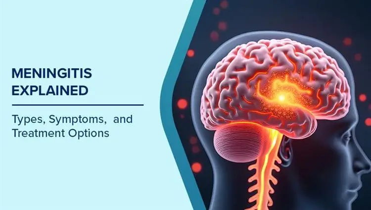
Category: Neurology | Medical Specialist: Neurologist; emergency physician for acute neurologic symptoms | References: American Academy of Neurology (AAN); WHO; National Institute of Neurological Disorders and Stroke (NINDS) Important: Meningitis can become life-threatening very quickly. If someone has a severe headache, fever, stiff neck, confusion, seizures, unusual sleepiness, or a rapidly developing non-blanching rash, seek emergency medical care immediately. Introduction: When a Headache Is More Than a Headache A headache is something most people have experienced. Meningitis is different. It is an inflammation of the protective membranes surrounding the brain and spinal cord, called the meninges. The condition can be caused by bacteria, viruses, fungi, parasites and, less commonly, certain medicines, cancers or injuries. The challenge is that early meningitis can look deceptively ordinary. Fever, headache, vomiting or tiredness may initially resemble flu or another common infection. But bacterial meningitis can deteriorate rapidly and requires urgent treatment. The World Health Organization (WHO) estimates that bacterial meningitis causes a particularly severe burden: approximately 1 in 6 people with bacterial meningitis die, while about 1 in 5 survivors develop serious long-term complications. That is why understanding meningitis isn’t simply about knowing a list of symptoms. It is about recognizing when an apparently routine illness has crossed into an emergency. What Is Meningitis? Meningitis occurs when the meninges—the protective layers around the brain and spinal cord—become inflamed. The inflammation may result from an infection or, less commonly, a non-infectious cause. Infectious meningitis is broadly classified according to the organism responsible. Major types of meningitis Type Common causes General pattern Treatment approach Bacterial meningitis Meningococcus, pneumococcus, Hib, Group B Streptococcus and others Often rapid and potentially life-threatening Urgent antibiotics and hospital care Viral meningitis Enteroviruses, herpesviruses and others Often less severe, but serious cases occur Supportive care; antivirals for selected infections Fungal meningitis Certain fungi, including Cryptococcus More common in people with weakened immunity Antifungal treatment Parasitic meningitis Certain parasites or amoebae Rare but potentially severe Cause-specific treatment Tuberculous meningitis Mycobacterium tuberculosis Often develops more gradually Prolonged anti-tuberculosis therapy The distinction matters because meningitis is not one disease with one treatment. Treatment depends heavily on its cause. What Causes Meningitis? 1. Bacterial infections Bacterial meningitis is the form doctors worry about most urgently. Important bacterial causes include: WHO identifies meningococcus, pneumococcus, Haemophilus influenzae and Group B Streptococcus as four major causes of acute bacterial meningitis globally. Some bacteria can live harmlessly in the nose or throat but occasionally invade the bloodstream or nervous system. 2. Viral infections Viral meningitis is the most common type of meningitis in the United States, according to the CDC. Enteroviruses are among the most common causes, while herpesviruses, influenza, mumps, measles and other viruses can also cause it. Many people with mild viral meningitis recover without specific treatment, but severe disease can require hospitalization. 3. Fungal and other causes Fungal meningitis is uncommon in healthy people but can be particularly serious in individuals with weakened immune systems. Other causes include certain parasites, amoebae, cancers, injuries and medications. How Does Meningitis Spread? There isn’t one universal transmission route. Some bacteria responsible for meningitis spread through respiratory droplets or throat secretions during close contact. Transmission risk depends on the organism and the circumstances. This is an important distinction: being exposed to a microorganism that can cause meningitis does not automatically mean a person will develop meningitis. For example, close contacts of someone with viral meningitis may acquire the virus but are not necessarily likely to develop meningitis themselves. For confirmed or strongly suspected meningococcal disease, however, certain close contacts may need preventive antibiotics. This should be determined by healthcare professionals or public-health authorities. Meningitis Symptoms: What Should You Watch For? The classic combination is: Fever + severe headache + stiff neck. But not everyone develops all three. Other symptoms can include: Symptoms may develop quickly, particularly in bacterial and meningococcal disease. Meningitis symptoms in babies Infants may not show the classic signs adults do. Watch for: Because babies cannot describe a headache or neck stiffness, behavioral changes can be especially important clues. The Meningitis Rash: An Important Red Flag A rash associated with meningococcal disease may appear as small red or purple spots that do not fade when pressed. However, there is a critical point to remember: Do not wait for a rash before seeking medical care. Not everyone with meningitis develops a rash, and symptoms can progress rapidly. The NHS specifically advises immediate medical attention when meningitis is suspected, even if not every symptom is present. Who Is at Higher Risk? Meningitis can affect anyone, but risk varies according to age, health status and exposure. Higher-risk groups can include: WHO notes that risk also varies according to geography, living conditions and the specific pathogen involved. How Is Meningitis Diagnosed? Meningitis cannot reliably be diagnosed from symptoms alone. A healthcare professional may use: Blood tests Blood samples can help identify infection and may allow laboratories to detect the responsible organism. Lumbar puncture A lumbar puncture, sometimes called a spinal tap, involves collecting cerebrospinal fluid (CSF) from the lower back. CSF analysis is central to diagnosing meningitis and can help distinguish bacterial, viral and other causes. WHO recommends performing lumbar puncture as soon as possible when appropriate, but emphasizes that suspected bacterial meningitis should not wait for the procedure before treatment begins. Other investigations Depending on the clinical situation, doctors may use: The goal isn’t simply to answer, “Does this person have meningitis?” It is also to determine what caused it, because that answer determines treatment. Treatment for Meningitis Treatment depends on the cause. Bacterial meningitis This is a medical emergency. When bacterial meningitis is suspected, antibiotics should be started as soon as possible. Treatment should not be postponed simply while waiting for lumbar puncture results or other investigations. Depending on the situation, doctors may also use intravenous corticosteroids, fluids, oxygen, seizure treatment and other supportive measures. The exact antibiotic regimen depends on factors such as age, suspected organism, immune status and local antimicrobial resistance patterns. Viral meningitis Many mild cases
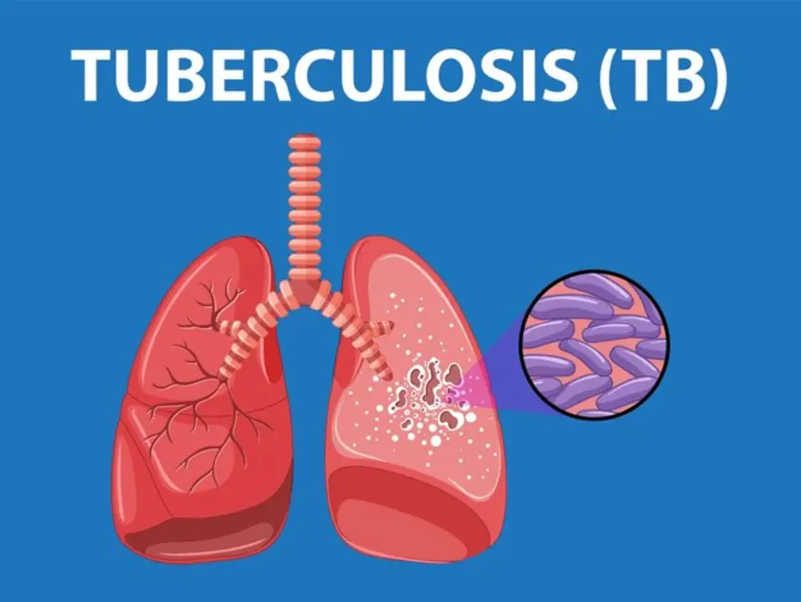
Category: Respiratory | Medical Specialist: Pulmonologist; primary-care physician; allergist where appropriate | References: Global Initiative for Asthma (GINA); Global Initiative for Chronic Obstructive Lung Disease (GOLD); WHO; CDC Introduction: Why Tuberculosis Still Matters A cough that refuses to go away can seem ordinary—until it isn’t. Tuberculosis (TB) remains one of the world’s most important infectious diseases, despite being preventable and curable. According to the World Health Organization (WHO), an estimated 10.7 million people developed TB in 2024, while about 1.23 million people died from the disease. India alone accounted for roughly 25% of global TB cases in 2024. What makes Tuberculosis particularly challenging is that it can be surprisingly quiet. Some people have symptoms for months before seeking care, while others have TB bacteria in their bodies without feeling ill at all. The encouraging part is equally important: TB can be diagnosed, treated and cured. The key is recognizing warning signs early, getting the right tests, and completing treatment exactly as prescribed. What Is Tuberculosis? Tuberculosis is an infectious disease caused by the bacterium Mycobacterium tuberculosis. It most commonly affects the lungs, where it is called pulmonary TB, but the bacteria can also affect other organs. TB spreads primarily through the air. A person with infectious pulmonary or laryngeal TB can release bacteria when coughing, sneezing, speaking or singing. Close, prolonged exposure—particularly in poorly ventilated spaces—can increase the chance of transmission. However, TB infection and TB disease are not the same thing. After someone inhales TB bacteria, the immune system may contain them. The bacteria can remain inactive, producing what is commonly called TB infection or latent TB. These people generally do not feel sick and do not spread TB. WHO estimates that approximately one-quarter of the world’s population has been infected with TB bacteria, but only about 5–10% of infected people eventually develop TB disease. TB Infection vs Active TB Disease Feature TB Infection Active TB Disease Bacteria present Yes Yes Symptoms Usually absent Often present Feeling ill Usually no Often yes Can spread TB? Generally no Pulmonary/laryngeal TB can be infectious Treatment Preventive treatment may be recommended Requires multidrug treatment Main concern Future progression to disease Illness and possible transmission This distinction is one of the most important things to understand about Tuberculosis. A positive TB infection test does not automatically mean that someone has contagious active TB. What Causes Tuberculosis? The immediate cause is infection with Mycobacterium tuberculosis. But developing active TB is influenced by more than exposure alone. The body’s immune defenses play a major role in determining whether bacteria remain contained or multiply and cause disease. Factors associated with a higher risk of developing TB disease include: WHO specifically identifies weakened immunity, diabetes, undernutrition, tobacco use and harmful alcohol use among important risk factors. A Useful Way to Think About TB Think of TB infection as a battle between the bacteria and the immune system. If the immune system successfully contains the bacteria, a person may remain well for years. If immune defenses weaken or the bacteria overcome containment, TB disease can develop. That’s why a person’s risk can change over time—even if the original exposure happened years earlier. Symptoms of Tuberculosis TB symptoms can develop gradually, which is one reason the disease may go unnoticed. Common Symptoms of Pulmonary TB Typical symptoms include: According to WHO, symptoms may be mild for many months. This delay can allow an infectious person to continue transmitting the bacteria before TB is recognized. TB Beyond the Lungs Tuberculosis does not always stay in the respiratory system. It can affect organs such as the: Symptoms therefore vary depending on the organ involved. For example, TB affecting the spine may cause persistent back pain, while TB involving the brain can produce neurological symptoms. Extrapulmonary TB can be harder to recognize because its symptoms may resemble many other conditions. When Should You See a Doctor? Don’t wait for symptoms to become severe. Medical evaluation is particularly important if you have a prolonged cough accompanied by symptoms such as unexplained weight loss, fever, night sweats, chest pain or blood in sputum. You should also discuss TB screening with a healthcare professional if you have had close contact with someone diagnosed with infectious TB or have medical or social factors that increase your risk. A cough alone does not prove that someone has TB—many other respiratory conditions can cause similar symptoms. The important point is that persistent or concerning symptoms deserve evaluation. How Is Tuberculosis Diagnosed? Diagnosis usually combines clinical assessment with laboratory and imaging tests. WHO recommends rapid diagnostic tests as the initial tests for people with signs and symptoms of TB. Molecular tests can help identify TB and, depending on the test, provide information relevant to drug resistance. Depending on the situation, healthcare professionals may use: A TB skin test or blood test can help identify TB infection, but it does not by itself establish that a person has active TB disease. Active disease must be evaluated separately. Tuberculosis Treatment: Why Completing the Course Matters One of the biggest mistakes people can make is stopping TB medication simply because they start feeling better. TB treatment uses multiple antibiotics, because the bacteria can be difficult to eliminate and can develop resistance when treatment is inadequate. For drug-susceptible TB, commonly used medicines include: WHO notes that treatment commonly lasts around 4–6 months, depending on the disease and regimen. Current treatment recommendations are periodically updated as evidence changes. The exact medicines, doses and duration should be determined by a qualified healthcare professional. Why TB Treatment Must Be Taken Seriously Imagine treating a fire by extinguishing only the visible flames. The remaining embers could reignite. TB bacteria can similarly persist when treatment is incomplete or inappropriate. Poor adherence can contribute to treatment failure and drug-resistant TB. That’s why modern TB care emphasizes not only medicines but also patient education, adherence support and follow-up. WHO’s current treatment guidelines specifically address both drug-susceptible and drug-resistant TB alongside patient care and support. Drug-Resistant Tuberculosis Some TB bacteria
Category: Cardiology | Medical Specialist: Cardiologist; emergency physician for acute symptoms | References: American Heart Association (AHA); American College of Cardiology (ACC); WHO Introduction: The Health Problem You May Not Feel Imagine feeling completely healthy, going to work, exercising occasionally, eating normally—and discovering during a routine check-up that your blood pressure is consistently high. There may be no pain, no obvious warning and no dramatic symptoms. That quiet nature is precisely what makes hypertension so important to understand. Hypertension, commonly called high blood pressure, occurs when the pressure of blood against artery walls remains persistently elevated. It affects an estimated 1.4 billion adults aged 30–79 worldwide, yet around 44% are unaware that they have it. Only about 23% are estimated to have their blood pressure under control. In other words, hypertension is not simply a number on a blood-pressure monitor. Over time, uncontrolled high blood pressure can place additional strain on the heart and blood vessels and increase the risk of stroke, heart disease, kidney disease and other complications. The encouraging part? Hypertension can often be prevented, treated and controlled. The first step is knowing your numbers and understanding what they mean. What Is Hypertension? Blood pressure is recorded using two numbers: WHO defines hypertension as blood pressure of 140/90 mmHg or higher, confirmed by measurements taken on two different days. However, blood-pressure categories and treatment thresholds can differ between clinical guidelines. The 2025 American Heart Association/American College of Cardiology guideline uses 130/80 mmHg as an important threshold for high blood pressure and sets an overall treatment goal of below 130/80 mmHg for many adults, with individual considerations for certain groups. Blood Pressure at a Glance Blood pressure reading General interpretation Below 120/80 mmHg Generally considered normal 120–129 and below 80 Elevated 130–139 or 80–89 High blood pressure under AHA/ACC classification ≥140 or ≥90 Hypertension under WHO definition >180 and/or >120 Severe hypertension requiring prompt assessment Classification systems vary, so your healthcare professional should interpret your readings in context. What Causes Hypertension? There isn’t always one obvious cause. For most adults, hypertension develops from a combination of genetic, biological, environmental and lifestyle factors. 1. Primary Hypertension Most hypertension is primary (essential) hypertension, meaning it does not have one identifiable underlying disease responsible for it. It tends to develop gradually as factors such as age, genetics, diet, body weight, physical activity and vascular changes interact over time. 2. Secondary Hypertension Sometimes high blood pressure is caused by another condition or medication. Possible contributors include: Treating the underlying cause can sometimes substantially improve blood pressure. Major Risk Factors for Hypertension Some risk factors cannot be changed, while others can. Non-Modifiable Risk Factors These include: Blood vessels naturally become less flexible with age, which can contribute to rising blood pressure. Modifiable Risk Factors This is where everyday choices can make a meaningful difference. Common contributors include: Some people are particularly sensitive to dietary sodium. Kidney disease, diabetes and metabolic syndrome can also increase vulnerability. A useful way to think about this is not “Did I cause my hypertension?”, but “Which factors can I change now?” That shift from blame to action can make hypertension much easier to manage. Symptoms of Hypertension: Why It Is Called a “Silent” Condition One of the biggest misconceptions about hypertension is that you will know when your blood pressure becomes high. Usually, you won’t. Many people with hypertension have no noticeable symptoms, even when their blood pressure has been elevated for a long time. Regular measurement is therefore much more reliable than waiting for warning signs. Some people may experience nonspecific symptoms such as: But these symptoms do not reliably diagnose hypertension. A headache, for example, does not automatically mean that your blood pressure is high. When High Blood Pressure Becomes an Emergency A reading above 180/120 mmHg requires immediate attention, particularly if it is accompanied by symptoms such as: These can indicate a hypertensive emergency or another serious medical problem. How Is Hypertension Diagnosed? A single elevated reading does not necessarily mean you have chronic hypertension. Healthcare professionals generally look for consistently elevated measurements, often using readings taken on separate occasions. Home blood-pressure monitoring can also help identify patterns that may not appear during a clinic visit. How to Get a More Accurate Reading Before measuring your blood pressure: Incorrect technique or cuff size can produce misleading results. Your clinician may also request blood and urine tests or other investigations to evaluate cardiovascular risk, identify possible secondary causes and determine whether organs such as the kidneys have been affected. Hypertension Treatment: What Actually Works? The best treatment plan depends on your blood-pressure level, overall cardiovascular risk, age, other health conditions and response to treatment. 1. Lifestyle Changes Lifestyle changes form the foundation of hypertension prevention and management. Eat for Your Blood Vessels Focus on: The DASH eating pattern is one evidence-based approach commonly recommended for blood-pressure management. Reducing heavily processed foods can be particularly useful because much of the sodium in modern diets comes from packaged and prepared foods rather than the salt shaker. Move More Regular physical activity can help lower blood pressure and improve cardiovascular fitness. You don’t necessarily need to transform your life overnight. Walking more, reducing prolonged sitting and gradually building consistent exercise habits can be more sustainable than an extreme short-term fitness plan. Maintain a Healthy Weight For people who are overweight, even modest weight loss may improve blood-pressure control and reduce cardiovascular risk. Limit Alcohol and Avoid Tobacco Excessive alcohol can contribute to elevated blood pressure, while tobacco damages blood vessels and increases cardiovascular risk. Prioritize Sleep and Stress Management Poor sleep and persistent stress can complicate blood-pressure control. Addressing sleep quality, maintaining a regular routine and using practical stress-management strategies can complement medical treatment. 2. Medicines for Hypertension Lifestyle changes are important, but they aren’t always enough. Depending on the individual situation, clinicians may prescribe medicines such as: The goal isn’t simply to take a tablet and see a lower number. Treatment aims to reduce long-term cardiovascular and organ
Category: General Medicine — Metabolic & Endocrine | Medical Specialist: Endocrinologist; primary-care physician; relevant subspecialist | References: American Diabetes Association (ADA); American Association of Clinical Endocrinology (AACE); Endocrine Society; WHO Introduction: Obesity Is More Than a Number on a Scale What if obesity had less to do with willpower—and much more to do with biology, environment, sleep, stress, medications, genetics and the world around us? That shift in perspective matters. Obesity is a chronic, complex disease involving excessive or abnormal fat accumulation that can affect nearly every major body system. The World Health Organization (WHO) describes obesity as a chronic, relapsing disease influenced by genetic, biological, behavioural, environmental and social factors. The scale can be useful, but it does not tell the whole story. Two people with the same weight may have very different health risks depending on where they carry fat, their metabolic health, activity levels and existing medical conditions. And obesity is becoming increasingly common. According to the WHO, 1 in 8 people worldwide were living with obesity in 2022, while adult obesity has more than doubled since 1990. Understanding obesity, therefore, is not about assigning blame. It is about identifying the factors involved and choosing an effective, sustainable treatment strategy. What Is Obesity? Obesity occurs when excess body fat accumulates to a level that may negatively affect health. For adults, body mass index (BMI) is commonly used as an initial screening measure: BMI in Adults General Classification Below 18.5 Underweight 18.5–24.9 Healthy range 25.0–29.9 Overweight 30 or above Obesity BMI is calculated from weight and height, but it is not a direct measurement of body fat. A muscular person, for example, may have a high BMI without having excessive body fat. Waist circumference and fat distribution can provide additional information about health risk. Recent clinical guidance also places greater emphasis on central adiposity—fat concentrated around the abdomen. NICE recommends considering waist-to-height ratio alongside BMI in appropriate adults because abdominal fat can provide additional information about cardiometabolic risk. A Better Question Than “How Much Do You Weigh?” Instead of looking only at weight, clinicians increasingly consider: This broader assessment helps transform weight management from a simple “lose weight” instruction into an individualized health plan. What Causes Obesity? There is rarely one single cause. A common explanation is that obesity develops when energy intake consistently exceeds energy expenditure. While this is biologically true, it is an incomplete explanation. WHO recognizes obesity as a multifactorial disease involving genetics, neurobiology, eating behaviours, environmental conditions, food availability and broader social factors. 1. Diet and Eating Patterns Frequent consumption of energy-dense foods and sugar-sweetened beverages can make it easier to consume more calories than the body needs. Large portions, frequent snacking, highly processed foods and liquid calories can contribute to gradual weight gain. The important point is that one meal does not cause obesity. Weight gain is usually the result of patterns repeated over weeks, months and years. 2. Physical Inactivity Modern lifestyles have reduced everyday movement. Desk-based work, long commutes, increased screen time and dependence on motorized transport can reduce energy expenditure. Exercise is not only about burning calories. Regular movement also supports cardiovascular health, muscle strength, insulin sensitivity, mood and sleep. 3. Genetics and Biology Genes can influence appetite, hunger signals, fat distribution, metabolism and how the body responds to weight loss. This helps explain why two people can follow seemingly similar lifestyles yet experience very different changes in weight. 4. Sleep and Stress Poor sleep can affect appetite regulation and food choices. Chronic stress may also influence eating behaviour, physical activity and hormonal pathways involved in weight regulation. A sustainable obesity-management plan should therefore look beyond the dinner plate. 5. Medical Conditions and Medications Some medical conditions can contribute to weight gain, while certain medications may also affect body weight. Examples can include some medicines used for mental health conditions, diabetes, seizures and other chronic diseases. This does not mean someone should stop a prescribed medicine because of weight gain. Instead, discuss concerns with a healthcare professional who can determine whether an alternative treatment is appropriate. 6. Environment and Social Factors Healthy choices are easier when healthy food is affordable and accessible, safe spaces for physical activity exist, and people have enough time and support to prioritize health. WHO emphasizes that obesity is influenced by the environments in which people live—not simply individual decisions. Symptoms of Obesity Unlike an infection or acute illness, obesity does not always produce a single obvious symptom. Some people may feel completely well while excess body fat is gradually increasing their risk of future disease. Possible signs or associated symptoms include: Importantly, not every symptom in a person with obesity is necessarily caused by their weight. NICE specifically warns clinicians against “diagnostic overshadowing”—assuming every health problem is caused by obesity and overlooking another diagnosis. Health Risks and Complications of Obesity Obesity can affect multiple systems rather than just appearance or body size. According to the National Institute of Diabetes and Digestive and Kidney Diseases (NIDDK), obesity is associated with conditions including type 2 diabetes, high blood pressure, heart disease, stroke, fatty liver disease, certain cancers, osteoarthritis, kidney disease, breathing problems and gallbladder disease. Area Affected Possible Complications Heart & blood vessels High blood pressure, heart disease, stroke Metabolism Insulin resistance, type 2 diabetes Liver Fatty liver disease Respiratory system Sleep apnea, breathing difficulties Musculoskeletal system Osteoarthritis, joint pain Digestive system Gallbladder and pancreatic problems Kidneys Increased risk of kidney disease Reproductive health Fertility and pregnancy-related problems Mental wellbeing Psychological distress and reduced quality of life The risk generally increases with the amount and distribution of excess body fat, although individual risk varies. Obesity vs. Being Overweight: What Is the Difference? The terms are related but not identical. Overweight generally refers to a BMI of 25 or higher in adults, while obesity generally begins at a BMI of 30. However, BMI is only a screening tool and should be interpreted alongside other clinical information. The comparison becomes especially important because health risks do not suddenly
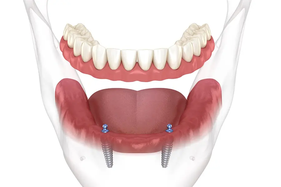
Category: Dental — Prosthodontics & Implants | Medical Specialist: Prosthodontist; implant dentist; oral and maxillofacial surgeon when indicated | References: American College of Prosthodontists (ACP); ADA; AAOMS Introduction: When “Loose Dentures” Become More Than an Annoyance A denture that moves when you chew, slips while you speak, or needs repeated adhesive can gradually change how you eat and socialize. For many people who have lost most or all of their teeth, the problem isn’t simply replacing missing teeth—it’s creating a prosthesis that feels secure enough to function naturally. That is where Overdentures can make a meaningful difference. Unlike conventional complete dentures, overdentures are designed to gain additional support or retention from remaining natural teeth, tooth roots, dental implants, or specially designed attachments. The result can be a more stable prosthesis without necessarily moving directly to a fixed implant-supported bridge. Recent research is particularly encouraging for implant-retained overdentures. A 2026 systematic review found that patients who transitioned from conventional complete dentures to implant-retained overdentures generally experienced improvements in satisfaction and oral-health-related quality of life. But are overdentures right for everyone? And how do they compare with conventional dentures? Let’s take a closer look. What Are Overdentures? An overdenture is a removable dental prosthesis that fits over and receives support from something beneath it. Depending on the patient’s oral condition, that support may come from: This makes overdentures different from conventional dentures, which depend primarily on the shape of the gums and underlying jawbone for support and retention. One of the most important distinctions is that an overdenture is not automatically an implant denture. Some overdentures are tooth-supported, while others are retained by implants. Why Do People Need Overdentures? The need for an overdenture usually develops from extensive tooth loss or difficulty obtaining adequate stability from conventional dentures. Common Causes 1. Extensive Tooth Loss When many teeth are missing, there may not be enough natural teeth remaining to support a conventional fixed restoration. An overdenture can replace the missing teeth while using selected remaining teeth or implants for additional support. 2. Poor Denture Retention Lower complete dentures are particularly prone to movement because the tongue, cheeks and lips constantly influence their position. An implant-retained overdenture can provide mechanical retention, reducing unwanted movement during everyday activities. Research has consistently investigated implant-retained mandibular overdentures as a way to improve retention and stability compared with conventional complete dentures. 3. Jawbone Resorption After tooth extraction, the alveolar bone gradually remodels and may shrink over time. This can make an old denture increasingly unstable. However, implant treatment is not simply about “stopping bone loss.” Treatment planning must consider existing bone volume, anatomy, systemic factors, prosthetic space and long-term maintenance. 4. Difficulty Eating or Speaking Repeated denture movement can make certain foods difficult to chew and may affect speech confidence. A more stable prosthesis can help patients regain confidence in everyday activities, although the degree of improvement varies from person to person. 5. Remaining Teeth That Can Still Be Used Sometimes the best solution isn’t to remove every remaining tooth. If selected teeth have a reasonable prognosis, they may be modified and incorporated into a tooth-supported overdenture. This approach can preserve natural tooth structures and provide additional proprioceptive feedback. That decision, however, requires careful periodontal, restorative and prosthodontic evaluation. Overdentures vs Conventional Dentures vs Fixed Implant Prostheses Choosing a prosthesis is less about finding the “best” option and more about finding the option that matches the patient’s anatomy, health, expectations, finances and ability to maintain it. Feature Conventional Denture Overdenture Fixed Implant Prosthesis Removable Yes Usually yes Usually no Natural teeth/root support No Sometimes No Implant support No Often Yes Stability Moderate to variable Usually improved Generally high Surgical treatment No May or may not require implants Usually required Cleaning Relatively simple Requires prosthesis and attachment care Requires meticulous home care Maintenance Relining/replacement may be needed Attachment and denture maintenance Implant and prosthetic maintenance Cost Generally lower Moderate to high Generally higher The important takeaway is that more expensive does not automatically mean more appropriate. For a patient with two healthy strategic teeth, a tooth-supported overdenture may be a sensible option. For someone with severe mandibular denture instability and suitable bone, an implant-retained overdenture may provide a substantial functional improvement. Types of Overdentures 1. Tooth-Supported Overdentures These are designed over retained natural teeth or prepared roots. The remaining teeth may receive special restorations or attachments that help support and stabilize the prosthesis. Potential advantages include: The downside is that the remaining teeth must be carefully selected and maintained. Root caries, periodontal disease and endodontic problems can compromise the treatment. 2. Implant-Retained Overdentures Implants can be placed in the jaw and connected to the removable denture using attachments. Common attachment designs include: There is no single attachment that is universally superior. A systematic review found broadly similar marginal bone-loss outcomes between ball and locator attachments, while also noting substantial variation among studies. 3. Mandibular Overdentures The lower jaw is a particularly important application because conventional mandibular dentures can be difficult to stabilize. Two-implant mandibular overdentures have long been an important evidence-based treatment approach, although newer research continues to examine alternatives such as single-implant and narrow-diameter implant designs. A 2026 systematic review found that single midline implant overdentures may be a suitable alternative for selected patients, but emphasized that current evidence is not strong enough to make them a universal replacement for two-implant protocols. Symptoms and Signs That May Suggest You Need More Denture Support Strictly speaking, overdentures do not have “symptoms” because they are a treatment rather than a disease. Instead, patients usually notice problems associated with inadequate denture support or tooth loss. Watch for: A loose denture isn’t necessarily a sign that you immediately need implants. Sometimes the issue is simply an old denture, inadequate fit, changes in the supporting tissues, or an incorrect bite. That is why a professional assessment matters. How Are Overdentures Diagnosed and Planned? The treatment begins with evaluation rather than simply choosing an attachment. A dentist or prosthodontist may assess: Oral Examination
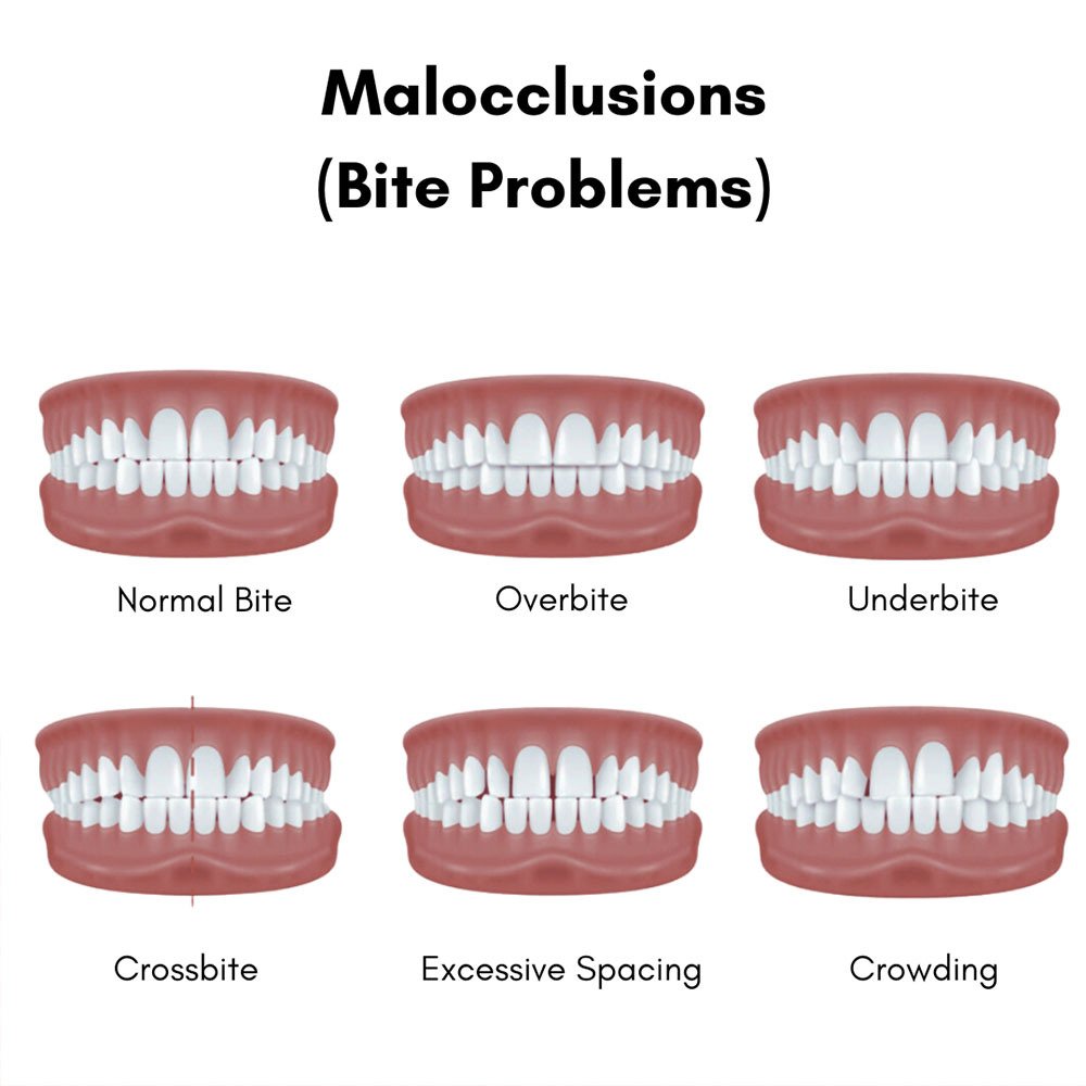
Category: Dental — Orthodontics & Occlusion | Medical Specialist: Orthodontist; dentist; oral and maxillofacial specialist when indicated | References: American Association of Orthodontists (AAO); ADA Introduction: When Your Bite Doesn’t Quite Fit Have you ever looked in the mirror and noticed crowded teeth, a prominent overbite, or a lower jaw that seems to sit farther forward? You may have wondered whether it is simply a cosmetic issue—or something that could affect your oral health. That is where Malocclusion comes in. Malocclusion describes an abnormal relationship between the upper and lower teeth or jaws when the mouth closes. In everyday language, it is often called a “bad bite.” It can range from mild crowding that causes little trouble to complex jaw discrepancies that affect chewing, speech, comfort, or dental health. Importantly, not every unusual-looking bite needs treatment. The real question is whether the bite is interfering with function, oral health, facial development, comfort, or the person’s goals. This distinction is one of the most useful things to understand before considering braces or clear aligners. What Is Malocclusion? Occlusion refers to the way the upper and lower teeth come together. Ideally, the teeth meet in a coordinated pattern that supports biting, chewing and speaking. Malocclusion occurs when the teeth or jaws do not relate to one another normally. It is not always obvious. A person may have visibly crooked front teeth but relatively good function, while another may have a less noticeable bite discrepancy that places excessive forces on certain teeth. In other words, straight teeth do not automatically mean a perfect bite—and a less-than-perfect smile does not automatically mean a serious problem. Common Types of Malocclusion Type What it means Common appearance Class I Back teeth have a relatively normal relationship, but crowding, spacing or other bite problems may exist Crooked or spaced teeth Class II Lower teeth or jaw are positioned behind the upper teeth/jaw Overjet or prominent upper front teeth Class III Lower teeth or jaw are positioned ahead of the upper teeth/jaw Underbite Overbite / Deep bite Upper front teeth overlap the lower front teeth excessively Lower front teeth partly covered Open bite Some teeth do not meet when the jaws are closed Gap between upper and lower teeth Crossbite One or more upper teeth bite inside or outside the corresponding lower teeth Teeth appear to “cross” Crowding There is insufficient space for teeth to align properly Overlapping or rotated teeth Spacing Excess space exists between teeth Noticeable gaps The American Association of Orthodontists (AAO) recognizes Class I, II and III relationships as important orthodontic classifications, while problems such as open bite, crossbite, overbite and overjet describe specific features of the bite. What Causes Malocclusion? Malocclusion rarely has one single cause. In many patients, several factors interact while the teeth and jaws are developing. 1. Tooth and Jaw Size Mismatch One of the most common underlying factors is a mismatch between tooth size and jaw size. For example, if the teeth are relatively large compared with the available jaw space, crowding can develop. Conversely, a relatively large dental arch or smaller teeth may contribute to spacing. 2. Genetics and Jaw Development The shape and growth pattern of the jaws are strongly influenced by genetics. A child may inherit a tendency toward a small upper jaw, a prominent lower jaw, or particular facial growth patterns. This is one reason orthodontic problems can appear to “run in families.” 3. Childhood Oral Habits Some habits can influence developing teeth and jaws, particularly when they persist over time. Examples include: Thumb sucking and tongue thrusting can place repeated forces on the developing teeth and contribute to problems such as increased protrusion or open bite. 4. Premature Loss of Baby Teeth Baby teeth do more than help children chew. They also help maintain space for permanent teeth. If a primary tooth is lost too early because of decay or trauma, neighboring teeth can drift into the space. That may reduce the room available for the permanent tooth that eventually needs to erupt. This is why treating childhood dental problems early can sometimes have orthodontic benefits later. 5. Missing or Lost Permanent Teeth Losing a permanent tooth can change the balance of the dental arch. Adjacent teeth may move into the empty space, while opposing teeth can shift vertically. Over time, this can contribute to a more complex bite. 6. Trauma and Developmental Conditions Jaw fractures, certain congenital conditions, and developmental differences can also affect tooth and jaw relationships. For these patients, orthodontic treatment may need to be coordinated with other dental or medical care. Symptoms of Malocclusion Some people notice Malocclusion immediately. Others live with it for years without realizing their bite is unusual. Possible signs and symptoms include: However, an important clinical point is worth emphasizing: jaw pain should not automatically be blamed on Malocclusion. The National Institute of Dental and Craniofacial Research notes that there is no evidence supporting the idea that malocclusion itself is a general cause of temporomandibular disorders (TMD). That means someone experiencing jaw pain deserves an appropriate evaluation rather than simply being told that their teeth need to be straightened. How Can Malocclusion Affect Oral Health? A poorly positioned tooth may be harder to brush and floss effectively. Crowded areas can therefore make plaque control more challenging. Depending on the individual’s bite, uneven forces may also contribute to abnormal tooth wear, fractures or mobility. MSD Manual notes that improperly distributed chewing forces can contribute to tooth wear and, in severe cases, damage to teeth and supporting structures. There can also be functional consequences. Severe Malocclusion may interfere with: This is why orthodontics is not simply about creating a straighter smile. The broader goal is to establish a healthy, functional bite. Diagnosing Malocclusion Diagnosis usually begins with a detailed dental and orthodontic examination. An orthodontist may evaluate: Depending on the situation, diagnostic records may include photographs, dental models or digital scans and X-rays. A panoramic X-ray can provide a broad view of the teeth and supporting structures,
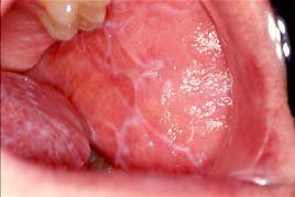
Category: Dental — Oral Medicine & Mucosal Conditions | Medical Specialist: Dentist; oral medicine specialist; dermatologist or physician when indicated | References: AAOM; ADA; NIDCR Introduction: When a “White Patch” Is More Than a White Patch A white, lace-like pattern inside the cheek can look harmless. Sometimes it is. But when those white lines persist, become sore, or appear alongside red, raw areas, they may point to a condition that deserves professional attention. Oral Lichen Planus (OLP) is a chronic inflammatory condition affecting the tissues inside the mouth. It can be painless and easy to overlook—or painful enough to make eating, brushing, and even speaking uncomfortable. The challenge is that it can resemble several other oral conditions, which is why recognizing the pattern is only the first step. The good news? Oral lichen planus can usually be managed. The goal is not simply to make a lesion “disappear,” but to control inflammation, relieve symptoms, identify possible triggers, protect oral health, and monitor persistent or changing areas over time. Important: Oral lichen planus should be diagnosed by a qualified dentist, oral medicine specialist, dermatologist, or physician. A persistent mouth lesion should not be self-diagnosed. What Is Oral Lichen Planus? Oral lichen planus is a chronic inflammatory disorder involving the oral mucosa. It is generally considered an immune-mediated condition in which immune cells, particularly cytotoxic T cells, attack cells within the oral epithelium. However, the exact trigger behind this immune response remains uncertain. Unlike a contagious infection, oral lichen planus cannot be passed from one person to another. It may occur on its own or alongside lichen planus affecting the skin or other mucosal surfaces. Interestingly, the mouth can sometimes be the only affected area. Common Areas Affected OLP may involve: The inside of the cheeks is particularly characteristic, especially when lesions appear symmetrically. What Causes Oral Lichen Planus? This is one of the frustrating aspects of OLP: there is usually no single identifiable cause. Researchers believe that an abnormal immune response plays the central role. Instead of protecting oral tissues, immune cells target epithelial cells, producing chronic inflammation. Several factors may contribute to or resemble OLP: 1. Immune-System Activity OLP is strongly associated with a T-cell-mediated inflammatory response. In many patients, however, the reason this response begins remains unknown. 2. Medications Some medicines can produce oral lichenoid reactions, which can look remarkably similar to oral lichen planus. Potentially associated medications include certain: This distinction matters because a medication-related lichenoid reaction may improve after the responsible drug is identified and appropriately changed under medical supervision. 3. Dental Materials and Contact Reactions In some cases, oral lichenoid lesions develop near dental restorations or in response to contact allergens. Materials such as metals, resins, or certain ingredients in oral-care products can occasionally be involved. That does not mean every person with OLP needs dental fillings removed. Removing restorations without evidence of a relevant contact reaction is unlikely to cure OLP. 4. Stress and Local Trauma Psychological stress and local mechanical irritation may aggravate existing lesions in some individuals. This is better understood as a potential trigger or exacerbating factor rather than a proven underlying cause. 5. Other Associations Hepatitis C has been associated with lichen planus in some populations, although the strength of this relationship varies geographically. Your clinician can decide whether additional medical evaluation is appropriate based on your individual history. Oral Lichen Planus Symptoms: What Does It Look and Feel Like? OLP does not look the same in everyone. Some people have no discomfort at all, while others experience significant burning and soreness. Common Signs and Symptoms Look out for: The classic white, branching lines are called Wickham striae. They are one of the visual clues clinicians may recognize during an oral examination. The Erosive Form Can Be More Painful Erosive oral lichen planus can produce red areas, superficial erosions, and ulceration. The gums may become particularly uncomfortable, sometimes producing a pattern known as desquamative gingivitis, where the surface tissue becomes red and peels away. This is where OLP can stop being a cosmetic concern and become a quality-of-life issue. Imagine trying to eat citrus fruit when the lining of your mouth is already inflamed. For someone with erosive OLP, that everyday experience can be surprisingly painful. Oral Lichen Planus vs Other Common Mouth Lesions Not every white or red oral lesion is OLP. Several conditions can look similar. Condition Typical appearance Pain Key consideration Oral Lichen Planus Lacy white lines, plaques, red/erosive areas Variable Often chronic and may be symmetrical Oral Thrush Creamy white patches Often burning Usually associated with Candida infection Leukoplakia Persistent white patch Often painless Requires professional evaluation Canker sore Localized ulcer Often painful Usually heals within a limited period Oral lichenoid reaction Similar to OLP Variable May relate to medication or contact exposure Oral cancer Persistent ulcer, lump, red/white lesion or other change May be painless Requires prompt assessment The important takeaway is simple: appearance alone cannot always establish the diagnosis. How Is Oral Lichen Planus Diagnosed? Diagnosis usually begins with a detailed oral examination and medical history. Your clinician may ask about: Why Is a Biopsy Sometimes Necessary? A biopsy may be recommended when the appearance is unusual, the diagnosis is uncertain, or an area needs to be evaluated for another condition. Microscopic examination can support the diagnosis and help distinguish OLP from other lichenoid conditions. Biopsy can also be important when clinicians need to rule out dysplasia or oral cancer. This is an important distinction: a biopsy does not mean your dentist thinks you have cancer. It is often a diagnostic safety step. Oral Lichen Planus Treatment: What Actually Helps? There is currently no guaranteed permanent cure for every case of OLP. Treatment is generally focused on controlling inflammation and symptoms while monitoring the condition. 1. Topical Corticosteroids For symptomatic oral disease, topical corticosteroids are commonly used and are considered a mainstay of treatment. They may be delivered as: More severe disease may require additional therapies. Because prolonged steroid use inside the mouth can
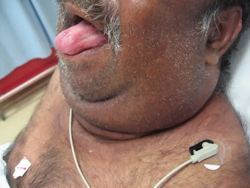
Category: Dental — Oral Surgery & Pathology | Medical Specialist: Oral and maxillofacial surgeon; oral medicine/oral pathology specialist; dentist | References: AAOMS; ADA; NIDCR Introduction: When a Tooth Infection Becomes an Emergency A toothache may seem like a problem that can wait until tomorrow. But when a dental infection begins spreading beneath the tongue and into the neck, the situation can change rapidly. Ludwig’s Angina is a rare but potentially life-threatening bacterial infection involving the floor of the mouth and the spaces beneath the jaw. Unlike a typical dental abscess, it can spread through interconnected tissue spaces and push the tongue upward, threatening the airway. That is what makes Ludwig’s Angina particularly important for both patients and dental professionals: the biggest danger isn’t simply the infection—it is the swelling that can interfere with breathing. Modern antibiotics, advanced imaging and better airway management have dramatically improved outcomes. Yet the condition still requires urgent medical attention because deterioration can occur quickly. Emergency warning: Difficulty breathing or swallowing, drooling, rapidly increasing swelling under the jaw or tongue, or a muffled/altered voice should be treated as an emergency. Do not wait for a routine dental appointment. What Is Ludwig’s Angina? Ludwig’s Angina is a rapidly spreading cellulitis, meaning inflammation and infection of soft tissues rather than a simple, well-contained abscess. It classically involves three connected anatomical spaces: The infection can cause firm swelling of these tissues and elevate the tongue. Because the tissues around the airway have limited room to expand, swelling can eventually make breathing difficult. The condition was first described in 1836 by German physician Wilhelm Friedrich von Ludwig. Despite its name, Ludwig’s Angina has nothing to do with the heart condition commonly called angina. What Causes Ludwig’s Angina? Dental infections are the leading cause One of the most important insights about Ludwig’s Angina is that a serious neck infection can begin with something as ordinary as an untreated dental infection. In particular, infections involving the lower second and third molars are strongly associated with Ludwig’s Angina. Current clinical references report that odontogenic infections account for the large majority of cases. Why do these teeth matter? The roots of lower molars can lie close to, or below, the attachment of the mylohyoid muscle. This anatomical relationship provides a pathway through which infection can spread into the submandibular tissues. Common dental sources include: Other possible causes Although dental infections dominate, Ludwig’s Angina can occasionally develop after: The infection is usually polymicrobial, meaning several types of bacteria may be involved. Aerobic and anaerobic organisms—including Streptococcus, Staphylococcus and anaerobic bacteria such as Bacteroides—can contribute. Who Is at Higher Risk? Ludwig’s Angina can occur in otherwise healthy people, but certain factors can increase susceptibility or make infection more difficult to control. Risk factors include: This is one reason preventive dental care matters beyond protecting teeth. Treating an infected tooth early can sometimes prevent a localized dental problem from becoming a deep neck infection. Ludwig’s Angina Symptoms: What Should You Watch For? Early symptoms can resemble an ordinary dental infection, which makes recognition especially important. Common symptoms Patients may experience: A characteristic finding is firm, bilateral swelling around the submandibular and submental areas, sometimes described as a “bull neck” appearance. The red flags matter most Not every symptom carries the same level of urgency. Symptom/sign What it may indicate Urgency Toothache with localized swelling Dental infection Prompt dental assessment Fever + jaw/neck swelling Spreading infection Urgent medical assessment Difficulty swallowing Increasing floor-of-mouth swelling Emergency Drooling Difficulty managing secretions Emergency Tongue elevation/swelling Airway displacement Emergency Muffled or altered voice Possible upper-airway involvement Emergency Difficulty breathing or stridor Threatened airway Immediate emergency care The evidence-based approach is straightforward: airway concerns take priority over diagnostic imaging or routine dental treatment. Why Ludwig’s Angina Can Become Dangerous So Quickly The anatomy of the floor of the mouth explains much of the danger. Think of the neck as a series of connected compartments. An infection does not necessarily remain where it started. It can travel along tissue planes, causing inflammation and swelling in neighboring spaces. As swelling increases, the tongue may be pushed upward and backward. The surrounding tissues can also become increasingly stiff and swollen. This creates a dangerous sequence: Dental infection → tissue-space spread → floor-of-mouth swelling → tongue elevation → airway narrowing → breathing difficulty That progression is why Ludwig’s Angina is considered a medical emergency rather than simply a severe dental infection. Ludwig’s Angina vs. a Typical Dental Abscess These conditions can start from the same dental problem, but their clinical behavior is different. Feature Typical Dental Abscess Ludwig’s Angina Main location Tooth/gum or localized tissue Floor of mouth and submandibular spaces Swelling Usually localized Often firm and more diffuse Airway risk Usually low Potentially life-threatening Tongue elevation Uncommon Characteristic concern Difficulty swallowing May occur Common Breathing difficulty Uncommon Major warning sign Treatment Dental source control ± antibiotics Airway management + IV antibiotics ± surgery The key lesson is that a dental infection should never be judged only by how painful the tooth feels. The location and direction of swelling can be much more important. How Is Ludwig’s Angina Diagnosed? Diagnosis is primarily clinical. A healthcare professional examines the mouth, tongue, jaw and neck while assessing the patient’s ability to breathe and manage secretions. Imaging A contrast-enhanced CT scan of the neck can help determine how far the infection has spread and whether an abscess has developed. However, imaging should not delay airway management in someone whose breathing is threatened. A patient who cannot safely lie flat or manage secretions may need immediate airway intervention rather than being sent for a scan first. Ultrasound may also be useful in selected situations, particularly when clinicians need additional information without immediately sending an unstable patient for CT. Ludwig’s Angina Treatment Treatment is typically carried out in a hospital and involves several teams, often including emergency medicine, anesthesiology, oral and maxillofacial surgery, ENT and critical care. 1. Protect the airway first This is the most important principle. If swelling threatens